Posted by Fred Koenig on May 3rd 2019
Dissecting Microscope Uses
A dissecting microscope (also known as a stereo microscope ) is called so because it is frequently used in dissecting operations. Its lower magnification ability, and long working distance range of 25 to 150 mm enables the user to manipulate the small specimen such as insects. A dissecting scope allows the user to observe live specimens as well as perform dissections under the microscope.
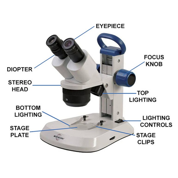
Parts of a Dissecting Microscope
Dissecting microscope parts include a single objective lens and two eyepiece lenses. There are two separate light paths transmitting the image of the specimen to each eyepiece. The beam of light comes from above the specimen. Since we have a binocular vision, our eyes see the image with overlapping field of view, which allows for the perception of depth. The result is an excellent three-dimensional image of the specimen under study.
Dissecting Microscope Applications
The dissecting microscope uses low magnification and offers a wide range of use. It is used in applications for the following fields:
Forensic Science
The long working distance (space between the specimen and objective lens), upright non-reversed image and wide field of view enable performing preliminary examinations of samples, manipulating small fibers or particles. It helps to prepare the samples for more detailed microscopical or instrumental comparisons or analyses. The long working distance and the fact that specimens are illumined by reflected light and rarely require any sample preparation is an added advantage. The samples can be directly placed under the forensic microscope and observed. It is useful to study bullet fragments, hair samples, surface markings, wires, fingerprints and many other details that are crucial for forensic reporting.
Gemology
It is used by experts in gems to determine the grade of the gemstones and for studying and evaluating gemstones. The wide field of view and lots of room between lens and stage makes it best suited for evaluating gemstones. For aspiring gemologists, 20X and 40X magnification is a good start but, if you want higher magnifications for future use, it is wise to find dissecting microscopes that offer higher magnification eyepieces for the model you want to purchase. For more, check out our gemological microscopes .
Surgical Applications
Dissecting microscope finds use in skin pathology applications. A dissecting scope is accurate in detecting basal and squamous cell carcinomas and in detecting a tumor in margins. The ability to visualize marginal surfaces in three dimensions allows for a thorough examination and quick localization and mapping of the involved tissues.
Schools and Labs
Dissecting microscopes are widely used by biology students to study insects and various samples.
So how different is the dissecting scope from that of a compound microscope, which is another widely used optical microscope? Besides the striking difference in the name, the two microscopes vary significantly in their parts and end use.
Compound vs. Dissecting Microscope
Dissecting microscopes are useful for observing the surface features of the specimen. On the other hand, a compound microscope is meant for looking through the specimen. Also, a compound microscope has a higher magnification ranging from 400X to up to 1000X while a dissecting microscope can magnify to a maximum of 70X.
If you are interested in procuring dissecting microscope for your school, laboratory or forensic uses, we have a wide selection from manufacturers. Call our toll free number 877.877.7274 or email info@nyscopes.com to get in touch with a microscopy expert to answer any queries on microscopes or orders.