Posted by Fred Koenig on Nov 20th 2020
Types of Microscopes
Microscopes come in a variety of sizes, styles and types in order to suit all of the many uses we have for them. Different technologies, quality levels, viewing results, and physical setups are required depending on what is being viewed, and for what reason.
Here is a comprehensive list of all types of microscopes:
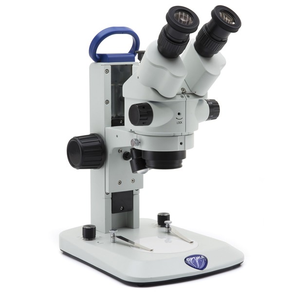
Stereo Microscopes
Stereo microscopes are named so because they employ more than one eyepiece, allowing the viewer to take advantage of our natural stereo vision – seeing things in 3D.
Stereo microscopes do not typically magnify things to the same degree as a compound microscope of the same quality level, but they do have the advantage of a greater working distance – allowing observation of larger objects without the need for slicing a sample for viewing. Magnification is typically only 10x to 40x. This lower magnification level, coupled with the larger field of view and working distance, allows much more manipulation of the object under observation. For opaque objects, both transmitted and reflected illumination are used (light from behind the object and above it, respectively), allowing for better 3D viewing.
For these reasons, stereo microscopes are often used for manufacturing things like circuit boards, for disdiv projects, and botanical observation and study. Excellent things to view under a stereo microscope include coins, flowers, insects and plant parts.
Read more about what a stereo microscope is here .
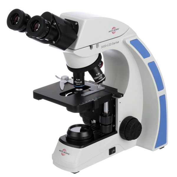
Compound Microscopes
A compound microscope (sometimes called a “biological microscope,” though this can technically include stereo microscopes as well) employs a single eyepiece and multiple “objectives” on a rotating ring which allows switching from one to another to increase magnification power. They are commonly seen in schools, especially at higher levels, and are widely used in medical research and other fields where a high degree of optical magnification is required.
Samples to be viewed must first be prepared on a slide, often with a cover slip to flatten the sample and keep it in place. Prepared slides are often available for student use, with samples permanently embedded between the layers.
Commonly viewed samples include blood and other human and animal cells (including cheek cells), parasites, bacteria, algae, and thin divs of other tissues and organs. These are not visible at all with the naked eye, and so are excellent candidates for viewing under a compound microscope.
Compound microscopes are most commonly able to magnify a range from 40x, through 100x, 400x and sometimes up as high as 1000x. Above that, and resolution drops dramatically, despite claims of higher magnification.
Read more about what a compound microscope is here .
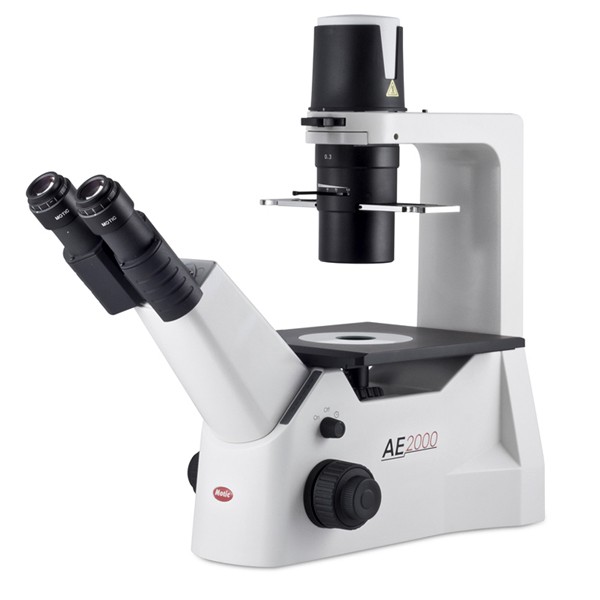
Inverted Microscopes
Inverted microscopes come in two common types: biological inverted microscopes and metallurgical inverted microscopes.
Biological inverted microscopes can usually magnify from 40x through 100x and sometimes as high as 200x or 400x. These are useful when observing living samples in petri dishes on a flat stage, with the objective lens housed below the stage. These are used commonly in industry and research around invitro fertilization, live cell imaging, developmental and cell biology, neuroscience, and microbiology.
Metallurgical inverted microscopes are commonly used, as the name would suggest, in metallurgical industry and research. They are used to find faults and fractures in metal objects and surfaces. Smooth, polished samples, which are commonly called “pucks,” are placed on the stage and viewing occurs through an objective housed beneath it.
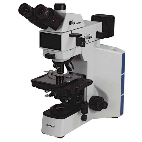
Metallurgical Microscopes
These high-powered microscopes are designed to assist in observing opaque samples - samples that do not allow light to pass through them. Reflected light illuminated the sample, which is then magnified from 50x to 100x, 200x and sometimes 500x its actual size. This allows observers to detect micron-level cracks in metals and paint, and to size substance grains.
Metallurgical microscopes are commonly used I the aerospace industry, in auto manufacturing, and any industry that deals with metals, glass, composites, crystals, polymers and ceramics.
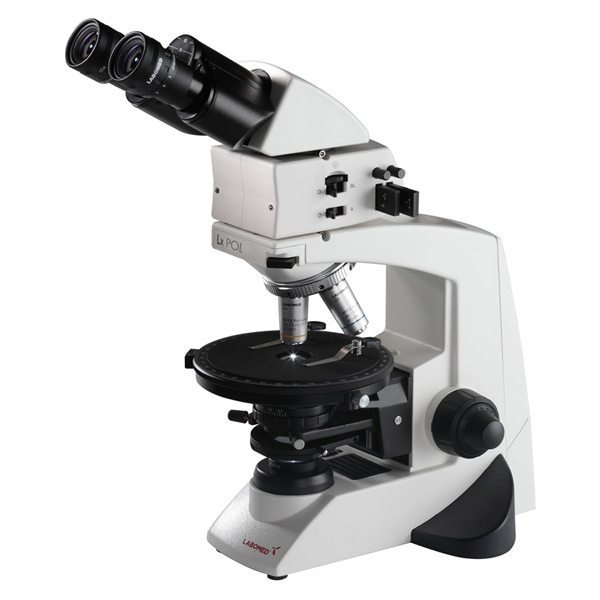
Polarizing Microscopes
Polarizing microscopes use light manipulation to increase the degree of contrast between different structures and densities under magnification. They use both transmitted and/or reflected light, filtered by a polarizer and controlled by the analyzer, to highlight differences in the texture, density and color of a sample surface. They are therefore excellent for viewing birefringent materials.
Polarizing microscopes are commonly used in geology, petrology, chemistry and many other similar industries.
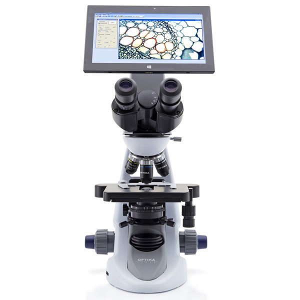
The Digital Microscope
Moving from the world of optical microscopes into the explosion of new images available through the use of digital microscopes , we can now see structures that are smaller than the wavelengths visible to the human eye. These were previously impossible to view, as wavelengths that we can see are simply too big to collide with the objects in a reliable and viewable frequency. Computers have changed all of this.
The digital microscope was invented in Japan in 1986. Some current models employ an eyepiece for viewing, though a computer monitor is more common. Software allows the user to focus on and record images (video and stills) of the sample under observation. Resulting files can be stored, sent, manipulated and used as any other digital image file.
Read more about what a digital microscope is here .
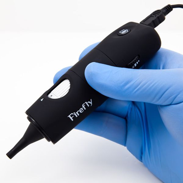
The USB Computer Microscope
USB computer microscopes can be used on almost any object, requires no preparation of the specimen, and is easy to use. It is basically a powerful macro lens (up to 200x) on the end of a USB cable. It has a small depth of field, but it can be a lot of fun and informative as a very powerful, digital magnifier.
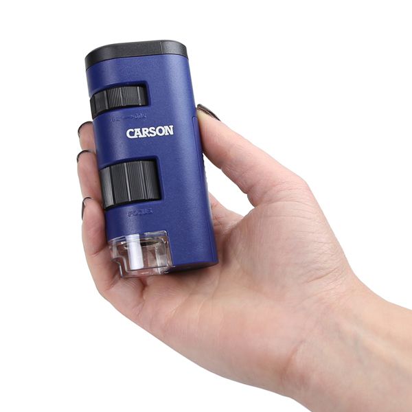
The Pocket Microscope
Another tiny option for the magnification enthusiast is the pocket microscope . This is also used by some scientists for hand-held imaging, especially when in the field. It can attain magnification levels of 25x to 100x, and there are some digital models now coming onto the market.
The Electron Microscope
The electron microscope (EM) is among the most powerful magnification tools available today.
The transmission electron microscope, or TEM, can display images of samples as small a 1 nanometer across. These are used in nanotechnology industries, and semiconductor analysis and production.
Scanning electron microscopes, or SEMs, are about 1/10 as powerful as a TEM. They can produce black and white and 3D images with sharp resolution.
Both TEMs and SEMs are widely used in biology, chemistry, gemology and metallurgy, and in other industries requiring microscopic topology, morphology and similar observations.
The Scanning Probe Microscope (SPM)
The scanning probe microscope, or SPM, has become a standard analysis tool in research and industry in the fields of physics, biology and chemistry.
Images are observed as highly magnified 3D images in real time – that’s the “scanning” aspect. The “probing” aspect is the device’s ability to probe the surface and draw out more information that static observation alone allows.
The Acoustic Microscope
Acoustic microscopes are a different type of microscope on a foundational level. Rather than striving to produce a visible image of a surface or object, acoustic microscopes seek to find faults, cracks and/or errors during the manufacturing process.
Through the use of high ultrasound, the microscope detects intra-cavity features very effectively. The most current form of it is called scanning acoustic microscopy (SAM). Internal structures can be viewed without staining or destroying the structure or the object surrounding it (if of a suitable size of course). Point focusing technology is employed to scan and penetrate the specimen while it is immersed in water, removing the need to damage it.
Confocal Microscope
The confocal microscope uses a laser to scan surfaces. Lasers do not require scanning mirrors, and can produce the data necessary to create a display image on a screen for detailed analysis.
Simple Microscope
This is the first kind of microscope that we know of, invented in the 17th century by a scientist named Antony van Leeuwenhoek. He combined a convex lens and a holding mechanism for specimens. His results were very modest by today’s standards of course, attaining a magnification maximum of around 300x. It was powerful enough, though, to illuminate a lot of previously unknown information, such as the presence of red blood cells of different shapes, the nature of cells in plan matter and other biological materials. It opened up a new field of study by extending the power of the human eye to see beyond what it developed to see on its own.
These microscopes are rarely used now, as the addition of a second lens – the part that changes a simple microscope into a compound microscope – we can greatly increase the power of magnification, without much additional cost or technology.
Do you need a New Microscope?
Here at New York Microscope Company, we stock thousands of microscopes for you to buy online today, you can shop our range here now .