Posted by Fred Koenig on Dec 16th 2020
Fluorescence Microscopy - Explanation and Labelled Images
How Does Fluorescence Microscopy Work?
A fluorescence microscope works by combining the magnifying properties of the light microscope with fluorescence emitting properties of compounds. Fluorescence microscopy uses a high-intensity light source that excites a fluorescent molecule called a fluorophore in the sample observed. The samples are labeled with fluorophore where they absorb the high-intensity light from the source and emit a lower energy light of longer wavelength. The resulting fluorescent light is then separated from the surrounding radiation with filters, allowing the observer to see only the fluorescing material. The resulting image is a magnified version of the specimen that is studied.
A majority of the fluorescence microscopes used in biology today are epi-fluorescence microscopes. Both the excitation and the observation of the fluorescence occur above the sample.
What is a Fluorescence Microscope?
A fluorescence microscope is used to study organic and inorganic samples. Fluorescence microscopy uses fluorescence and phosphorescence to examine the structural organization, spatial distribution of samples. It is particularly used to study samples that are complex and cannot be examined under conventional transmitted-light microscope. Fluorescence microscopy images helps to study substances present in low concentrations where high-sensitivity is crucial to detect them.
The Characteristics of a Fluorescence Microscope
The main parts of a fluorescent microscope overlap with the traditional light microscope. However, there are two main features that sets fluorescent microscope apart from the traditional microscope. One is the type of light source and the other is the use of specialized filter elements. A fluorescence microscope uses a higher intensity light to illuminate the samples.
Parts of a Fluorescence Microscope
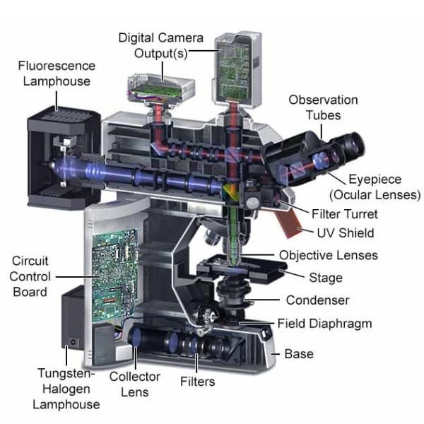
A powerful light source (xenon or mercury arch lamp): The light emitted from the mercury arc lamp is 10-100 times brighter than most incandescent lamps and provides light in a wide range of wavelengths, from ultra-violet to the infrared.
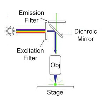
Excitation filter: The purpose of the excitation filter is to filter out all wavelengths of the light source, except for the excitation range of the fluorophore under inspection. The brightness and brilliance of images are dictated by the minimum transmission percentage of the filter. The ideal transmission being >85%.
The Dichroic mirror (beamsplitter): The Dichroic mirror or beamsplitter is placed at an angle of 45° between the excitation filter and emission filter. The function of a dichroic filter is to reflect the excitation signal towards the fluorophore and to transmit the emission signal towards the detector.
Emission filter: The emission filter is located within the imaging path of a fluorescence microscope. Its job is to filter out the entire excitation range and to transmit the emission range of the fluorophore under inspection.
Objective lens: The purpose of the objective lens is to transmit light to the sample to form the image. The light passes down through the dichroic mirror before reaching the objective lens.
Camera system: A camera system helps to record the images of the specimen with high-resolution. CCD (Charge Coupled Device) cameras are often used in the system. These electron multiplying cameras are good at imaging single photon events without loss of sensitivity. They do not require an image intensifier, and images can be captured at high speed.
What Are Fluorescence Microscopes Used For?
Fluorescence microscopy finds use in biology, biomedical and material sciences. The unique functionality of fluorescence microscopes helps identify cells and sub-microscopic cellular components with accuracy and details.
Fluorescence microscopy uses are widely used in the field of histochemistry to detect particles such as neurotransmitter amines which cannot be seen by conventional microscopes.
It is used in food chemistry to assess the presence, structural organization and spatial distribution of specific food components in a product.
Another use of fluorescence imaging is Fluorescence Speckle Microscopy. It is a technology that uses fluorescence labeled macromolecular assemblies such as cytoskeletal protein to study movement and turnover rates.
Fluorescence microscopy staining also is helpful in the field of mineralogical applications. It is routinely used for the study of minerals such as coal, graphene oxide and more.
It is also widely used in the textile industry to analyze fiber dimensions. Epifluorescence microscopy helps to study the fiber-based materials including paper and textiles.
Fluorescence microscopy is ideal for studies of porosity in ceramics, using a fluorescent dye. It is also applicable to studies of semiconductors.
Light Sources in Fluorescence Microscopy
Fluorescence microscopy uses intense levels of near-monochromatic illumination, and therefore requires one of four main types of lamp: xenon arc lamps or mercury-vapor lamps (with an excitation filter), supercontinuum sources, high-powered LEDs, or lasers. Lasers are the most common choice for confocal microscopy, total internal reflection fluorescence microscopy , and other complex techniques. In cases where widefield epifluorescence microscopes are used, xenon lamps, mercury lamps, or LEDs with a dichroic excitation filter are more appropriate. Two micro lens arrays in the illumination path of a widefield epifluorescence microscope can produce an illumination coefficient of variation between 1 and 2%, with high uniformity.
You can shop our range of LED fluorescence microscopes here .
Fluorescence Sample Preparation
It goes without saying that a sample must be fluorescent to be suitable for fluorescent microscopy, but a few words on preparing samples for viewing might be useful. More than one method exists, and the most common ones include labelling with fluorescent stains, expression of a fluorescent protein, or taking advantage of the intrinsic fluorescence (autofluorescence) of a sample. Fluorescence microscopy is a common and powerful tool in the life sciences.
Epifluorescence microscopy
In epifluorescence microscopy, the light of the excitation wavelength passes through the objective lens to illuminate the specimen. The same objective focuses the fluorescence to the detector. This objective lens generally has a higher numerical aperture for greater resolution. Most of the light is transmitted through the specimen, so only reflected excitatory light passes through to the objective, along with the emitted light. The epifluorescence method produces a higher ratio of signal to noise, producing a better image to the viewer.
Wavelengths are filtered by the dichroic beam splitter, which reflects the unneeded excitation light back toward the source, but allows the fluoresced light through to the viewer.
You can buy epifluorescence microscopes here .
Biological Fluorescent Stains
Some stains are useful for a wide range of biological substances. Some small molecules have their own fluorescence and a preferred biological molecule with which to bond. Examples include DAPI and Hoechst, which are excited by UV light; and DRAQ5 and DRAQ7, which excite best under red light. Each of these binds to the minor groove of DNA, and by doing so, highlight (or “label”) the nuclei of cells.
Some other stains are peptides, toxins and drugs, and bind to specific cellular structures. These must first be derivatized with a fluorescent reporter, in order to work. Phalloidin is a good example of this type of fluorescent stain. It is used primarily to stain actin fibers in the cells of mammals. Collagen Hybridizing Peptide is another, newer one, that can be used on denatured collagen fibers. Stains and dyes that bind cellulose or pectin are good candidates for the staining of plant cell walls. Numerous fluorescent molecules, called fluorophores or fluorochromes, can link (chemically) to different molecules in order to bind them to a target of interest within a sample. Fluorescein, Alexa Fluors, and DyLight 488 are examples of these.
Even with this array of excellent fluorescent probes, researchers are constantly seeking new and more specific candidates, especially those that allow live imaging of plant cells.
Immunofluorescence
The highly specific binding of an antibody to its antigen, thereby labelling specific proteins or other molecules within the cell, is achieved through a process known as immunofluorescence. This process begins with the treating of a sample with a primary antibody specific to the molecule the observer wishes to label. Sometimes this is achieved by conjugating a secondary antibody to a fluorophore that binds to the first antibody, resulting in the same labelling. A primary antibody in a mouse, for example, might recognize tubulin. If this is combined with a secondary anti-mouse antibody derivatized with a fluorophore, an observer could label microtubules in a cell.
Fluorescent Proteins
Modern genetic knowledge and techniques allow scientists to modify DNA. The same techniques can be used to genetically modify proteins to add a fluorescent protein reporter, which means that scientists can thereby make a certain protein fluorescent. Once it is fluorescent, it can be directly tracked, even in living cells.
Further Reading
You can learn more about fluorescence microscopy below:
Sources:
http://www.scholarpedia.org/article/Fluorescent_proteins
https://pubmed.ncbi.nlm.nih.gov/18228363/
https://www.ncbi.nlm.nih.gov/pmc/articles/PMC4711767/